Total Price: $119.95
Contact Information
Foxp3/Transcription Factor Staining Kit
SKU: E-CK-A108-20
Foxp3/Transcription Factor Staining Kit
Introduction
Elabscience® Foxp3 / Transcription Factor Staining Kit has been formulated and optimized for staining with antibodies to transcription factors and nuclear proteins, such as Foxp3 and STAT3.
Instructions
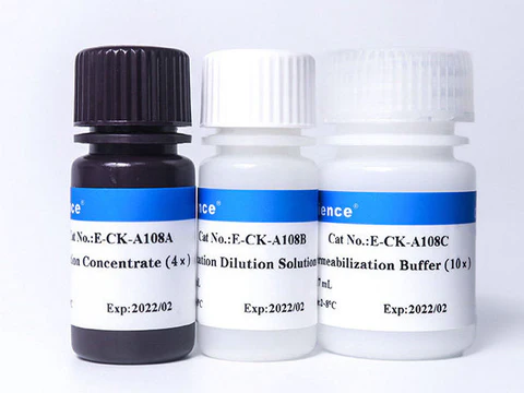
- Dilute Fixation Concentrate (4×) with Fixation Dilution Solution to 1× Fixation Working Solution before use.
- For example, take 1 mL Fixation Concentrate (4×) [E-CK-A108A] and add it to 3 mL Fixation Dilution Solution [E-CK-A108B] to get 4 mL 1× Fixation Working Solution. Each sample requires 1 mL of 1× Fixation Working Solution.
- Dilute Permeabilization Buffer (10×) with ddH2O to 1×Permeabilization Working Solution before use.
- For example, take 1 mL Permeabilization Buffer (10×) [E-CK-A108C], and add it to 9 mL ddH2O to get 10 mL 1× Permeabilization Working Solution. Each sample requires 6.5 mL of 1× Permeabilization Working Solution.
Experimental Procedure
- Add the single-cell suspension into tubes, 1×10⁶ cells in 100 μL suspension per tube.
- [Optional] Stain cells with a Fixable Viability Dye.
- [Optional] Block Fc receptors in cell suspensions according to experimental requirements.
- Stain cell surface markers. Refer to the FCM protocol (Staining Cell Surface Targets for Flow Cytometry).
- After incubating with the cell surface marker, add 1 mL of Cell Staining Buffer [E-CK-A107], centrifuge samples at 300×g for 5 min, discard the supernatant, then resuspend the cells with 100 µL of Cell Staining Buffer [E-CK-A107].
- Add 1 mL of 1×Fixation Working Solution to each tube and mix fully, incubate the cells at 4°C for 30 min, then centrifuge at 600×g for 5 min and discard the supernatant.
- Add 2 mL of 1×Permeabilization Working Solution to each tube and mix fully, centrifuge at 600×g for 5 min and discard the supernatant.
- Repeat Step 7.
- Resuspend the cells with 100 µL of 1×Permeabilization Working Solution.
- Without washing, add the recommended amount of directly FCM antibody for detection of intracellular antigen(s) to cells and incubate for at least 30 min at room temperature in the dark.
- Add 2 mL of 1×Permeabilization Working Solution to each tube and centrifuge at 600×g for 5 min at room temperature. Discard the supernatant.
- Resuspend the cells with appropriate Cell Staining Buffer [E-CK-A107], then analyze the samples by flow cytometer.
Storage
Store at 2~8°C for six months in the dark. Avoid freeze / thaw cycles
Notes
- It is normal for the Permeabilization Buffer (10×) to have precipitation, and it will not affect the use effect.
- For maximal assay performance, this reagent should be used within 6 months. Avoid freeze / thaw cycles.
- The fixation and permeabilization steps that are required for the detection of intracellular antigens may alter the light scatter properties of cells and may increase non-specific background staining. Including extra proteins such as BSA or fetal calf serum (FCS) in the staining buffer may help reduce non-specific background. The use of Fixable Viability Dyes is recommended to help eliminate dead cells during the analysis.
- For your safety and health, please wear the lab coat and disposable gloves before the experiments.