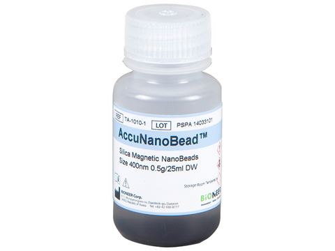
Bioneer AccuNanoBead Ni-NTA Magnetic Beads, Size 400nm (0.5 g/25 ml)
SKU: TA-1017-1
Bioneer AccuNanoBead™ Ni-NTA Magnetic Beads, Size 400nm (0.5 g/25 ml)
Buy Bioneer products from MSE Supplies at the best value.
Magnetic beads are used as materials for cell experiments, DNA purification and disease observation, and can also be used as materials for direct treatment. This AccuNanoBead™ have the advantages of high Binding Capacity due to their relatively large surface area because they are nano-sized.
Recently, there have been many researches applying magnetic beads in the field of biology: from cellular experiments and DNA purification to disease monitoring and even disease treatment. The surface of magnetic beads are coated with functional groups that allows binding of DNA, RNA, proteins, and specific cells. Afterwards, the beads are moved or attached to a desired position through external magnetic fields to perform purification. In addition, while conventional Magnetic Beads are micro-sized, this AccuNanoBeads™ is nano-sized, bearing the advantages of high binding capacity due to their relatively large surface area.
Features and Benefits
- Miniscule size: Average sizes of 400 nm magnetic nanobeads
- Uniformed size distribution
- Sphere form of silica coated magnetic nanobeads
- Excellent protein purification efficiency
- Rapid purification
Applications
- Magnetic DNA/RNA Purification
- Magnetic Protein Purification
- Magnetic Biosimilar Separation
- Magnetic Antibody Separation
Specifications
Protein Purification Method by using Magnetic Nanobeads
After loading the protein sample to the tube, add the magnetic beads to combine them. Afterwards, fix the beads by using external magnetic field and wash the solution. Finally, elute the DNA or RNA from the magnetic beads. This method provides faster DNA purification than traditional ones.
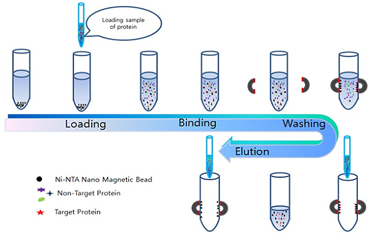
Figure 1. Recombinant Protein and Antibody protein purification protocol by Magnetic Nanobeads.
FE-SEM and TEM Images of Magnetic Nanobeads
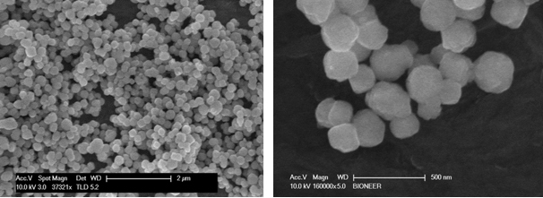
Figure 2. FE-SEM picture of Magnetic Nanobeads. The electron microscope picture shows spherical magnetic nanobeads.
EDS Analysis of Magnetic Nanobeads

Figure 3. EDS analysis results of Magnetic Nanobeads. The results show that the Magnetic Nanobeads consist of silica and iron oxide.
Size Distribution of Magnetic Nanobeads

Figure 4. Particle size distribution of Magnetic Nanobeads. The average size of the Magnetic Nanobeads is about 400 nm.
Comparison of Protein Purification Beads
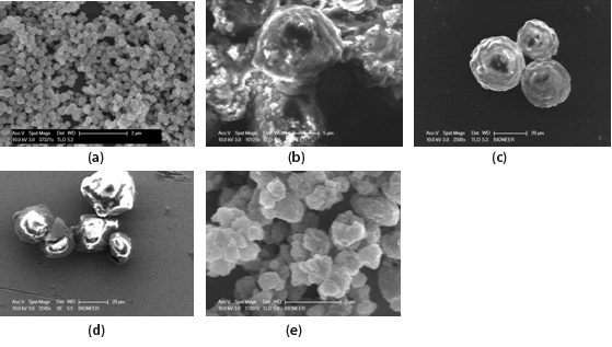
Figure 5.
(a) SEM image of 450 nm Bioneer Magnetic Nanobeads
(b) SEM image of A company Magnetic Beads
(c) SEM image of B company Magnetic Beads
(d) SEM image of C company Magnetic Beads
(e) SEM image of D company Magnetic Beads
Figure 5 shows scanning electron microscope images of magnetic beads from Bioneer and a different company (b~e). Scanning electron microscope image of synthesized magnetic nanobeads shows that magnetic nanobeads of Bioneer is 400 nm size and spherical shape. However, the magnetic beads from the other company are micro meter size and various shape.
Protein Purification Yield
Table 1 shows the protein purification yield of Bioneer Ni-NTA magnetic nanobeads. From these results, it can be seen that Bioneer magnetic nanobeads have a high protein purification yield of 77%. The protein used in the experiment was a 17kDa His-Tagged protein. The yield is the amount of target protein purified from the loading protein.

Table 1. Protein purification yield of bioneer Ni-NTA magnetic nanobeads (30 mg).
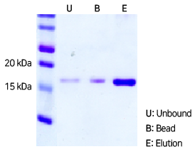
Figure 6. SDS-PAGE image of purified 17 kDa His-tagged protein with Bioneer Ni-NTA magnetic nanobeads.
