Mouse PDGF-AA(Platelet Derived Growth Factor AA) ELISA Kit
SKU: E-EL-M3079
Mouse PDGF-AA(Platelet Derived Growth Factor AA) ELISA Kit
| Detection Range | 0.78-50 pg/mL |
| Sensitivity | 0.47 pg/mL |
Product Details
Properties
| Assay type | Sandwich-ELISA |
| Format | 96T |
| Assay time | 3.5h |
| Detection range | 0.78-50 pg/mL |
| Sensitivity | 0.47 pg/mL |
| Sample type &Sample volume | serum, plasma and other biological fluids; 100μL |
| Specificity | This kit recognizes Mouse PDGF-AA in samples. No significant cross-reactivity or interference between Mouse PDGF-AA and analogues was observed. |
| Reproducibility | Both intra-CV and inter-CV are < 10%. |
| Application | This ELISA kit applies to the in vitro quantitative determination of Mouse PDGF-AA concentrations in serum, plasma and other biological fluids. |
Test Principle
This ELISA kit uses the Sandwich-ELISA principle. The micro ELISA plate provided in this kit has been pre-coated with an antibody specific to Mouse PDGF-AA. Standards or samples are added to the micro ELISA plate wells and combined with the specific antibody. Then a biotinylated detection antibody specific for Mouse PDGF-AA and Avidin-Horseradish Peroxidase (HRP) conjugate are added successively to each micro plate well and incubated. Free components are washed away. The substrate solution is added to each well. Only those wells that contain Mouse PDGF-AA, biotinylated detection antibody and Avidin-HRP conjugate will appear blue in color. The enzyme-substrate reaction is terminated by the addition of stop solution and the color turns yellow. The optical density (OD) is measured spectrophotometrically at a wavelength of 450 nm ± 2 nm. The OD value is proportional to the concentration of Mouse PDGF-AA. You can calculate the concentration of Mouse PDGF-AA in the samples by comparing the OD of the samples to the standard curve.
Kit Components and Storage
An unopened kit can be stored at 2-8℃ for 1 month. If the kit is not supposed to be used within 1 month, store the components separately according to the following conditions once the kit is received.
| Item | Specifications | Storage |
|---|---|---|
| Micro ELISA Plate(Dismountable) | 96T: 8 wells ×12 strips 48T: 8 wells ×6 strips |
-20℃, 6 months |
| Reference Standard | 96T: 2 vials 48T: 1 vial |
|
| Concentrated Biotinylated Detection Ab (100×) | 96T: 1 vial, 120 μL 48T: 1 vial, 60 μL |
|
| Concentrated HRP Conjugate (100×) | 96T: 1 vial, 120 μL 48T: 1 vial, 60 μL |
-20℃(Protect from light), 6 months |
| Reference Standard & Sample Diluent | 1 vial, 20 mL | 2-8°C, 6 months |
| Biotinylated Detection Ab Diluent | 1 vial, 14 mL | |
| HRP Conjugate Diluent | 1 vial, 14 mL | |
| Concentrated Wash Buffer (25×) | 1 vial, 30 mL | |
| Substrate Reagent | 1 vial, 10 mL | 2-8℃(Protect from light) |
| Stop Solution | 1 vial, 10 mL | 2-8°C |
| Plate Sealer | 5 pieces | |
| Manual | 1 copy | |
| Certificate of Analysis | 1 copy |
Technical Data:
Precision
Intra-assay Precision (Precision within an assay): 3 samples with low, mid range and high level Mouse PDGF-AA were tested 20 times on one plate, respectively.
Inter-assay Precision (Precision between assays): 3 samples with low, mid range and high level Mouse PDGF-AA were tested on 3 different plates, 20 replicates in each plate.
| Intra-assay Precision | Inter-assay Precision | |||||
|---|---|---|---|---|---|---|
| Sample | 1 | 2 | 3 | 1 | 2 | 3 |
| n | 20 | 20 | 20 | 20 | 20 | 20 |
| Mean (pg/mL) | 2.33 | 7.11 | 18.26 | 2.16 | 7.69 | 19.09 |
| Standard deviation | 0.15 | 0.31 | 0.77 | 0.13 | 0.44 | 1.02 |
| CV (%) | 6.28 | 4.40 | 4.20 | 5.96 | 5.76 | 5.34 |
Recovery
The recovery of Mouse PDGF-AA spiked at three different levels in samples throughout the range of the assay was evaluated in various matrices.
| Sample Type | Range (%) | Average Recovery (%) |
|---|---|---|
| Serum(n=8) | 94-109 | 100 |
| EDTA plasma (n=8) | 94-107 | 101 |
| Cell culture media (n=8) | 92-103 | 98 |
Target Information
| Synonyms | PDGF-AA |
| Research Area | Cancer, Cardiovascular, Metabolism, Immunology, Signal transduction |
Assay Procedures
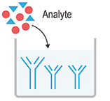 |
1. Add 100μL standard or sample to the wells. Incubate for 90 min at 37°C |
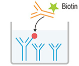 |
2. Discard the liquid, immediately add 100μL Biotinylated Detection Ab working solution to each well. Incubate for 60 min at 37°C |
 |
3. Aspirate and wash the plate for 3 times |
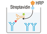 |
4. Add 100μL HRP conjugate working solution. Incubate for 30 min at 37°C. Aspirate and wash the plate for 5 times |
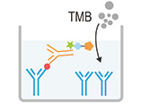 |
5. Add 90μL Substrate Reagent. Incubate for 15 min at 37°C |
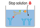 |
6. Add 50μL Stop Solution |
 |
7. Read the plate at 450nm immediately. Calculation of the results |


